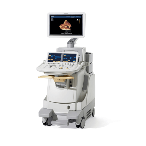
Combining unprecedented 2D and 3D image quality in the same transducer and a host of easy-to-use quantification, clinical performance, and information management tools, the new iE33 xMATRIX echo system addresses the clinical needs of managing patients with cardiac disease, including heart failure, valvular disease, and congenital heart disease.
Philips iE33 Echocardiography Ultrasound
Description
Versatile X5-1 transducer
With 3,000 elements and breakthrough PureWave xMATRIX technology, the X5-1 supports virtually any cardiac ultrasound exam, including 3D, 2D, color flow, M-mode, PW/CW Doppler, Tissue Doppler imaging, and contrast-enhanced exams. It delivers outstanding image quality and a new handle design with relaxed grip to reduce user fatigue and improve scanning stability.
Acquire crisp, high-resolution 2D images, even on your most difficult patients.
Use iRotate electronic rotation to more easily obtain challenging views, such as apical 2 chamber, and to reduce foreshortening.
Switch from 2D to 3D imaging at the touch of a button to quickly integrate volume imaging into routine exams.
Image the entire heart in 3D, in real time with Live Volume, and enlarge and rotate volumes with iCrop.
Reduce your stress in stress echo
The new Philips iE33 xMATRIX automates stress echo exams, so they’re fast and consistent. Use iRotate Stress Echo in combination with the X5-1 transducer to complete an entire stress echo protocol. You can include acquisition of 2-chamber, 3-chamber, and 4-chamber 2D images all from the standard windows following peak patient exertion—without rotating the transducer.
As you move electronically around the heart capturing baseline images, making any necessary small angle adjustments, these adjustments will be saved, along with gain and depth settings, for instant recall when you are in your post-exercise or -pharmacologic stage. A much less stressful stress echo solution that benefits both you and your patients.
With CMQ-Stress, you can quantify 2D stress echo studies and communicate global and regional LV function during each stage. And Live 3D stress echo with iSlice lets you add volume acquisition to your stress protocol and slice it later for the best views and content to make informed diagnoses—with confidence.
Accurate 2D and 3D LV function and regional wall motion assessment
Cardiac Motion Quantification (CMQ) increases the accuracy of LV function and wall motion measurement. The new CMQ speckle tracking algorithm makes strain analysis a useful tool in assessing presence and extent of LV disease includes multiple strain and strain rate parameters, including longitudinal strain.
Cardiac 3D Quantification Advanced (3DQ Advanced) is the first semi-automated analysis of true LV volumes.
3DQ Advanced uses all the voxels available to generate a full 3D endocardial border with greater accuracy.
Waveform display provides accurate data for assessing global function based on LV volume, ejection fraction and stroke volume. Simultaneous display of 17 regional waveforms enables temporal comparisons between segments.
More information for interventional procedures
Live 3D TEE (transesophageal echo) is providing clinical cardiologists, cardiac surgeons, anesthesiologists, interventional cardiologists, and echocardiographers with views of cardiac structure and function seen for the first time. Capturing a 3D volume is quick, accurate, reproducible, and quantifiable. You can view the 3D heart in real time as well as review the 3D volume at any time, select any plane for interrogation, and perform extensive quantification. And now, with the new dual volume display, you can open a volume data set like a book and see inside or view anatomy from opposing sides simultaneously, as well as perform distance and area measurements directly on the volume. It’s more information for diagnosis and treatment planning.
Award-winning service
Philips support services are designed to maximize uptime. Our Remote Services connectivity allows for many advanced service features, including virtual on-site visits for both clinical and technical support. It provides faster resolution to issues and questions, remote clinical education, and remote log file transfer to minimize downtime.
We offer Remote Desktop, “over the shoulder” system monitoring for faster technical and clinical troubleshooting and training options. And to help you manage your practice, we provide utilization reports with system and exam data analysis.

